Guess the Spot (not where you think)

Jammer

❤2Hike
.
Joined
Feb 23, 2012
Messages
655
OK -- it's not my turn in Guess the Spot, but I REALLY wanted to post this one. This can be a one-off picture instead of a new thread. I just liked this picture because it's a VERY familiar spot, but I never would have guessed it (not my picture.) I realize it's difficult because it's such a CLOSE shot, but any guesses where this is:
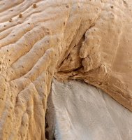

WII_5.jpg

- Jamal
 
It is a SEM (scanning electron microscope) shot of the eye to eyelid interface on a bed bug.
 
Haha my first thought was finger nail, but it looked so worn and beat up that I thought it might be from one of your feet after one of your epic treks :)
 
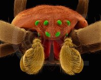
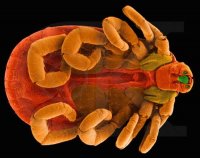
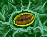
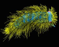
Hey, we were right, or close to it. I love SEM photos. In a former life I used a SEM for investigations and did a bit of photomicrography. Here are some favorites (not my work). I did work on metals. These pics are animals under a SEM colorized to bring out details...

sem_1.jpg

sem_2.jpg

sem_3.jpg

sem_4.jpg
 
Hey, we were right, or close to it. I love SEM photos. In a former life I used a SEM for investigations and did a bit of photomicrography. Here are some favorites (not my work). I did work on metals. These pics are animals under a SEM colorized to bring out details...

Really!!! That's what I currently do. Actually most of my research has been in TEM but have done a fair amount of SEM including cryo-SEM. Our lab was the first to take images of snowflakes in an SEM. Right now I have to go look at some Salmonella.

http://emu.arsusda.gov/snowsite/
 
Balto,

The snow crystal SEM photomicrographs... That's you ?!?! They are great. I love the crystalography they display. Makes my metals work look simplistic. I am an engineer and hired a SEM lab to help me with forensics on failure causes in adhesive bonding in the construction of gyroscopes and lasers for inertial navigation systems. Most of my optical photomicrography was for similar reasons. I always admired the people that were able to do SEM work on objects (snowflakes) that required keeping the object of interest in much lower than room temperatures. Must be a pain in the @#@#@. Have you done any scanning tunneling microscopy? Maybe that is what you mean by TEM. I will be sure to chase down some of your work through the link you provided. Thanks!

BTW give us a pic of a salmonella bacteria in your "chamber".. (hands wringing)
 
@ Art: Salmonella was negatively stained, looks like a black sperm. Not very exciting. The snow pictures I linked were from our lab but not my images. A former scientist was our snow guru, Eric Erbe. Google his name, USDA microscopy and snow for other images. He was probably the best cryo-microscopist ever.
No scanning tunneling for me. TEM means Transmission Electron Microscopy. Fixed biological tissue, ultra thin section it, stain it then view on a TEM. All of my projects are biological. Too many to mention but if it affects our agriculture products then I am looking at it. The parasites are by far the coolest to investigate. I am lucky to have such an interesting job. Now if I can do this part time and play more all would be golden.
 
Back
Top